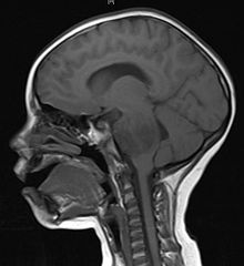
RESEARCHES GOING ON BRAIN TUMOR
Google images
Vesicular stomatitis virus
In 2000, researchers at the University of Ottawa, led by John Bell PhD., discovered that the vesicular stomatitis virus, or VSV, can infect and kill cancer cells, without affecting healthy cells if coadministered with interferon.
The initial discovery of the virus' oncolytic properties were limited to only a few types of cancer. Several independent studies have identified many more types susceptible to the virus, including glioblastoma multiforme cancer cells, which account for the majority of brain tumors.
In 2008, researchers artificially engineered strains of VSV that were less cytotoxic to normal cells. This advance allows administration of the virus without coadministration with interferon. Consequently administration of the virus can be given intravenously or through the olfactory nerve. In the research, a human brain tumor was implanted into mice brains.
Research on virus treatment like this has been conducted for some years, but no other viruses have been shown to be as efficient or specific as the VSV mutant strains. Future research will focus on the risks of this treatment, before it can be applied to humans.[24]
Retroviral replicating vectors
Led by Prof. Nori Kasahara, researchers from USC, who are now at UCLA, reported in 2001 the first successful example of applying the use of retroviral replicating vectors towards transducing cell lines derived from solid tumors. Building on this initial work, the researchers applied the technology to in vivo models of cancer and in 2005 reported a long-term survival benefit in an experimental brain tumor animal model. Subsequently, in preparation for human clinical trials, this technology was further developed by Tocagen, Inc. (Toca 511 & Toca FC) and is currently under clinical investigation in a Phase I/II trial for the potential treatment of recurrent high grade glioma including glioblastoma multiforme (GBM) and anaplastic astrocytoma.[25]
Identification of a Cancer Stem Cell in Human Brain Tumors
Most current research on human brain tumors is focused on the molecular and cellular analysis of the bulk tumor mass. However, there is overwhelming evidence in some malignancies that the tumor clone is heterogeneous with respect to proliferation and differentiation. In human leukemia, the tumor clone is organized as a hierarchy that originates from rare leukemic stem cells that possess extensive proliferative and self-renewal potential, and are responsible for maintaining the tumor clone. They report here the identification and purification of a cancer stem cell from human brain tumors of different phenotypes that possesses a marked capacity for proliferation, self-renewal, and differentiation. The increased self-renewal capacity of the brain tumor stem cell (BTSC) was highest from the most aggressive clinical samples of medulloblastoma compared with low-grade gliomas. The BTSC was exclusively isolated with the cell fraction expressing the neural stem cell surface marker CD133. These CD133+ cells could differentiate in culture into tumor cells that phenotypically resembled the tumor from the patient. The identification of a BTSC provides a powerful tool to investigate the tumorigenic process in the central nervous system and to develop therapies targeted to the BTSC.[26]
Combination of Chemotherapy and Radiation Therapy Lengthens Lives of Patients With Anaplastic Oligodendroglial Tumors
A recent study by the European Organisation for Research and Treatment of Cancer (EORTC) shows that chemotherapy after radiation therapy slowed the growth of anaplastic oligodendroglial tumors (a type of brain tumor). It also lengthened the lives of patients with this type of tumor, especially for those whose tumor was missing specific genetic material in chromosomes 1 and 19 (called 1p/19q co-deletions). Currently, most patients with this disease receive either chemotherapy or radiation therapy, but not both.
In this study, 368 patients with newly diagnosed anaplastic oligodendroglial tumors who had not received treatment were given either radiation therapy alone or radiation therapy plus chemotherapy. Chemotherapy was given in six cycles or rounds with the drugs procarbazine (Matulane), lomustine (CeeNu), and vincristine (Vincasar).
For patients receiving the combination of chemotherapy and radiation therapy, the time it took for the disease to worsen was about two years, compared with a little over a year for patients receiving only radiation therapy. In addition, patients receiving the combination treatment lived with their disease for about a year longer than those who received only radiation therapy. About 80 patients in this study had a 1p/19q co-deletion. These patients who received radiation therapy and chemotherapy were about half as likely to die from the disease as those who received radiation therapy.[27]
New Tumor Marker Affects Prognosis in Glioma
Researchers have discovered a correlation between a tumor protein called epidermal growth factor receptor (EGFR) vIII ("variant three") and the prognosis (chance of recovery) of grade 3 and 4 primary brain tumors called gliomas. Primary brain tumors begin in the brain. A grade 4 tumor, also called glioblastoma, is the fastest-growing brain tumor. Grade 3 tumors are moderately aggressive, but may grow as fast as grade 4 tumors in some patients.
In this study, doctors studied 63 tumor samples from patients with grade 3 tumors. They found that patients whose tumor samples had the vIII protein lived an average of 7.2 months, whereas the patients whose tumors did not have vIII lived an average of 33 months.
Jan Buckner, MD, lead author and Professor of Oncology at the Mayo Clinic College of Medicine in Rochester, Minn, explained that this information could be used to identify which grade 3 gliomas might grow as quickly as grade 4 gliomas. "vIII expression is characteristic of glioblastoma multiforme, the most aggressive primary brain tumor. It is reasonable to treat patients with grade 3 tumors and vIII expression as glioblastoma patients." These results confirm previous findings that genetic markers of glioblastoma in grade 3 tumors are associated with a poor prognosis.
"Eventually, vIII expression may be important in selecting patients for clinical trial participation and for therapies that target individual molecular profiles," said Dr. Buckner.[28]
PET imaging of brain tumor with [methyl-11C]choline.
This describes a new method of [11C]choline synthesis for intravenous injection. Aimed at the utilization of this compound for brain tumor imaging with PET. METHODS: After [11C]carbon dioxide production in a cyclotron and the subsequent [11C]methyl iodide synthesis, [methyl-11C]choline was synthesized by the reaction of [11C]methyl iodide with "neat" dimethylaminoethanol at 120 degrees C for 5 min. Purification was achieved by evaporation of the reactants followed by passage of the aqueous solution of the product through a cation-exchange resin cartridge. The time required for overall chemical processing, excluding the cyclotron operation, was 15 min. Radiochemical yield was > 98%. Radiochemical purity was > 98%. Chemical purity was > 90% (dimethylaminoethanol was the only possible impurity). Specific radioactivity of the product was > 133 GBq/mumol. The whole body distribution was examined in rabbits with PET. Clinical studies were performed in patients with brain tumor using PET after intravenous injection of 370 MBq of [11C]choline.
RESULTS: In rabbits,[11C]choline was taken up from blood by various tissues very rapidly, and the radioactivity remaining in blood became almost negligible 5 min after intravenous injection. Taking advantage of this characteristic, we obtained stable tissue distribution images of human brain using PET. In patients with brain tumor, PET produced clearly delineated positive images of the tumors. CONCLUSION: Carbon-11-choline can be used for obtaining clear images of brain tumor in PET[29]
